 |
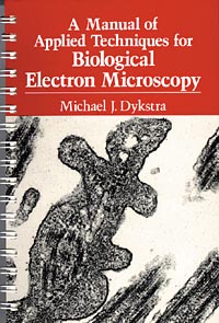
A Manual of Applied Techniques for Biological Electron Microscopy
Michael J. Dykstra,
1993, 2nd Printing, 272pp, spiral bound,
ISBN 03064-44496.
Currently Out Of Print |
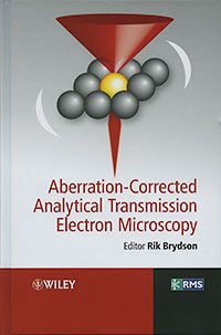 |
Abberation-Corrected Analytical Transmission Electron Microscopy
Rik Brydson, editor
2011, 280pp, hardcover,
ISBN 978-0-470-51851-9.
Electron microscopy has undergone significant developments in recent years due to the practical implementation of schemes which can diagnose and correct for the imperfections (aberrations) in both the probe-forming and the image-forming electron lenses. This book presents the background and implementation of techniques which have allowed true imaging and chemical analysis at the scale of single atoms as applied to the fields of materials science and nanotechnology.
Edited and written by the founders of the world's first aberration corrected Scanning Transmission Electron Microscope facility (SuperSTEM at Daresbury Laboratories in the UK), this text:
- Presents the theory, instrumentation and applications of aberration correction in transmission electron microscopes
- Is based on an established course taught at postgraduate summer schools by leaders in this field
- Is essential reading for researchers involved in the analysis of materials at the nanoscale
|
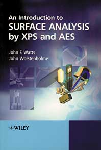 |
An Introduction to Surface Analysis by XPS and AES
John F. Watts, John Wolstenholme
May 2003, 224pp, paperback,
ISBN 0-470-84713-1.
An Introduction to Surface Analysis by Electron Spectroscopy is a clear and
accessible introduction to the key spectroscopic techniques used in surface
analysis. Focusing on the two most popular surface science techniques; X-ray
photoelectron spectroscopy (XPS) and Auger electron spectroscopy (AES), the
book will be of benefit to both students and users in industry who require a
rapid grounding in the methods before carrying out their own analysis.
|
|
Starting with an introduction to the basic concepts of electron spectroscopy, the text moves on
to explain underlying physical principles, discusses the instrumentation employed and looks at the
interpretation of the resulting spectra. The latest material on angle resolved XPS, surface engineering
and complimentary methods have been included to provide an up-to-date account of these widely used techniques.
- Examples taken from the fields of metallurgy, corrosion, microelectronics, polymers and adhesion.
- Detailed glossary of key surface analysis terms.
- An invaluable text for undergraduate and postgraduate students studying surface analysis within
science and engineering.
- A useful reference to those working within the field and needing to familiarize themselves with
these important techniques.
|
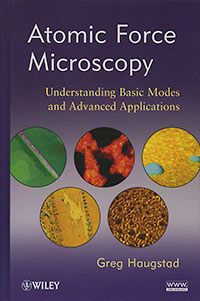 |
Atomic Force Microscopy: Understanding the Basic Modes and Advanced Applications
Greg Haugstad
2012, 464pp, hardcover,
ISBN 978-0-470-63882-8.
Although Atomic Force Microscopy (AFM) is an essential tool in materials and biological research, little systematic training is available for users. Addressing the gap in the field, Atomic Force Microscopy is a comprehensive primer covering knowledge readers need in order to become astute operators of AFM, including basic principles, data analysis, and such applications as imaging, materials property characterization, in-liquid interfacial analysis, tribology (friction/wear), electrostatics, and more.
|
Geared to a wide audience, from students to technicians to research scientists and engineers, this unique guide explains in simple terms the distance-dependent intersurface forces AFM users need to understand when measuring basic surface properties. Moving gradually to more complex areas, it explores such topics as calibration, physical origins of artifacts, and multifrequency methods. Features include:
- Emphasis on core methods available on most research-grade commercial systems including ancillary modes such as lateral force probes or interleave-based scanning
- Clarification of essential concepts needed for using dynamic AFM and examining phase images
- Examples of simple yet useful custom methods to enable shear modulation and setpoint ramping
- A companion website containing real AFM data files and theoretical constructs for analyzing data
Readers will learn to configure and operate instruments and interpret results for successful applications of atomic force microscopy. They will also gain a thorough understanding of a variety of topics for future research and experimentation. |
 |
Confocal Microscopy Methods and Protocols
Stephen W. Paddock, editor
442pp, hardcover,
ISBN 0896035263.
Stephen Paddock and a highly skilled panel of experts lead the researcher using
confocal techniques from the bench top, through the imaging process, to the journal
page. They concisely describe all the key stages of confocal imaging-from tissue
sampling methods, through the staining process, to the manipulation, presentation,
and publication of the realized image. Written in a user-friendly, nontechnical style,
the methods specifically cover most of the commonly used model organisms: worms, sea
urchins, flies, plants, yeast, frogs, and zebrafish. The powerful hands-on methods
collected here will help even the novice to produce first-class cover-quality confocal
images.
|
 |
Conn’s Biological Stains, 10th Edition
R.W. Horobin & J.A. Kiernan, editors
2002, 538pp, hardcover,
ISBN 1859960995.
|
 |
Dictionary of Microscopy
Julian P. Heath
2005, 358pp, softcover,
ISBN 0-470-01199-8.
Provides concise definitions of over 2,500 terms used in the fields of light
microscopy, electron microscopy, scanning probe microscopy and X-ray microscopy
and related techniques. Written by Dr. Julian P. Heath, Editor of Microscopy and
Analysis. This dictionary is intended to provide easy navigation through the microscopy
terminology and to be a first point of reference for definitions of new and established
terms.
|
 |
Electron Microscopy and Analysis, 3rd Edition
Peter J. Goodhew, John Humphreys, Richard Beanland,
2000, 272pp, softcover,
ISBN 0-748-40968-8.
The text is liberally illustrated and now incorporates questions and answers with each chapter.
Already established laboratory manual, this edition will be essential reading for both the
materials scientist and the bio-scientist.
Table of contents: Microscopy with Light and Electrons; Electrons and Their Interactions with
the Specimen; Electron Diffraction; The Transmission Electron Microscope; The Scanning Electron
Microscope; Chemical Analysis in the Electron microscope; Electron Microscopy and Other Techniques;
Further Reading; Index.
|
 |
Electron Microscopy Methods and Protocols
M. A. Nasser Hajibagheri, editor
1999, "Methods in Molecular Biology", v 117, spiral bound,
ISBN 0-896-03640-5.
See all chapters for this book
|
 |
Electron Microscopy, 2nd Edition
John J. Bozzola & Lonnie D Russell
1999, 688pp, hardcover,
ISBN 0-763-70192-0.
Covers all of the important aspects of electron microscopy for biologists, including theory
of scanning and transmission, specimen preparation, digital imagine and image analysis,
laboratory safety and interpretation of images. The text also contains a compete atlas of
ultrastructure. This new edition contains 200 additional line drawings and micrographs. Both
scanning and transmission microscopes are covered at an introductory level, eliminating the
need for students to buy multiple books. Includes and excellent chapter on safety in the electron
microscopy lab. Thought provoking questions for each chapter appear at the end of the book.
|
 |
Electron Probe Quantitation
K.F.J. Heinrich & Dale E. Newbury, editors
1991, 400pp, hardcover,
ISBN 0-306-43824-0.
|
 |
Fundamentals of Light Microscopy and Electronic Imaging
Doulgas B. Murphy,
2002, 378pp, hardcover,
ISBN 0-471-23429-X.
Presents the fundamentals of light microscopy, including the basics of microscope design,
image formation, and camera function. Researchers will learn how to acquire electronic images
and perform image processing. Provides instructions on the latest techniques involving confocal
laser scanning microscopy, digital CCD microscopy, as well as a chapter covering issues related
to image processing for scientific publication.
|
 |
Handbook of Sample Preparation for Scanning Electron Microscopy and X-Ray Microanalysis
Patrick Echlin,
2009, 330pp, hardcover,
ISBN 978-0-387-85730-5.
|

|
Immunogold-Silver Staining: Principles, Methods and Applications
M. A. Hayat
1995, 336pp, hardcover,
ISBN 0-849-324491.
|
|
In Situ Hybridization in Electron Microscopy
Gerard Morel, Annie Cavalier, Lynda Williams
2001, 472pp, hardcover,
ISBN 0-849-30044-4.
|
 |
Introduction to Focused Ion Beams: Instrumentation, Theory,
Techniques and Practice
Lucille A. Giannuzzi, editor,
358pp, softcover,
ISBN 978-1441935748.
Introduction to Focused Ion Beams is geared towards techniques and applications. This is the only text that discusses and presents the theory directly related to applications and the only one that discusses the vast applications and techniques used in FIBs and dual platform instruments.
|
 |
Introduction to Immunocytochemistry, 3rd Edition
J. M. Polak & S. Van Noorden,
179pp, softcover,
ISBN 1859962084.
|
|
Methods in Plant Electron Microscopy and Cytochemistry
William V. Dashek, editor
2000, 312pp, spiral bound,
ISBN 0-896-03809-2.
|
|
Microscopy and Histology for Molecular Biologists: A User’s Guide
J.A. Kiernan & I. Mason, editors
2002, 400pp,
hardcover, ISBN 1-85578-141-7;
paperback, ISBN 9781855781566.
|
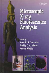 |
Microscopic X-Ray Fluorescence Analysis
Koen H. A. Janssens, Freddy C. V. Adams, Anders Rindby, editors
June 2000, 434pp, hardcover,
ISBN 0-471-97426-9.
In the last 10 - 15 years many analytical advances in X-ray fluorescence analysis (XRF) have taken place,
giving rise to non-destructive ultrasensitive surface analyses of materials. One of the variants of XRF
developed is micro-XRF (µ-XRF), which is able to analyze the distribution of major, minor and trace elements
in microscopic sample surface areas. Due to the availability of commercial instrumentation and the development
of simple devices for focusing X-rays, µ-XRF has increased in popularity and is able to fill the gap between
bulk X-ray fluorescence and electron probe X-ray microanalysis (EPXMA).
|
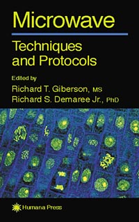 |
Microwave Techniques and Protocols
Richard T. Giberson & Richard S. Demaree, Jr., editors
2001, 300pp, hardcover,
ISBN 0-89603-903-X.
This is the first book to provide detailed protocols from laboratories that integrate
microwave technology as part of their normal routine. The text shows how microwave
processing has evolved into a methodology that greatly reduces processing times without
sacrificing quality, and can be utilized by any laboratory personnel who process samples
for microscopy. This volume includes examples of technicians and investigators using light
microscopy tissue samples in paraffin, plastics, as well as in vitro labeling and immuno
labeling. Also included are cases demonstrating the processing of samples for scanning and
transmission electron microscopy. Microwave Techniques and Protocols sets the trend by
assembling a wide range of detailed protocols that are being used today to improve productivity
and reduce costs.
|
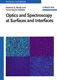 |
Optics and Spectroscopy at Surfaces and Interfaces
Vladimir G. Bordo, Horst-Günter Rubahn
December 2005, 282pp, paperback,
ISBN 3-527-40560-7.
This book covers linear and nonlinear optics as well as optical spectroscopy at solid surfaces and at
interfaces between a sold and a liquid or gas. In the first part, the author gives a concise introduction
to the physics of surfaces and interfaces. They discuss in detail physical properties of solid surfaces and
of their interfaces to liquid and gases. The necessary theoretical background for understanding various optical
techniques is provided thereafter.
|
|
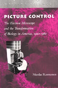
Picture Control: The Electron Microscope and the Transformation of
Biology in America, 1940-1960
Nicolas Rasmussen,
1999, 436pp, paperback,
ISBN 0-804-73850-5.
|
|
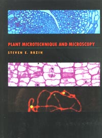
Plant Microtechnique and Microscopy
Steven E. Ruzin,
1999, 336pp, spiral bound,
ISBN 0-19-508956-1.
|
|
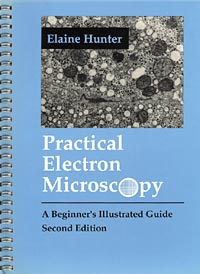
Practical Electron Microscopy, A Beginner’s Illustrated Guide,
2nd Edition
Elaine Hunter,
1993, 173pp, spiral bound,
ISBN 0-521-38539-3.
|
|
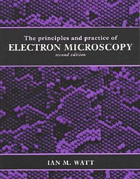
The Principles and Practice of Electron Microscopy, 2nd Edition
Ian M. Watt,
1997, 596pp, softcover,
ISBN 0-521-43591-9.
|
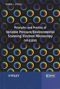 |
Principles and Practice of Variable Pressure/Environmental Scanning Electron Microscopy (VP-ESEM)
Debbie J. Stokes
2008, 221pp, hardcover,
ISBN 978-0-470-06540-2.
Scanning electron microscopy (SEM) is a technique of major importance and is widely used throughout the scientific and technological communities. A recent development is the advent of SEMs that utilise a gas for image formation while simultaneously providing the charge stabilisation for electrically non-conductive specimens and/or a suitable environment for materials and experiments involving water and other gases.
The book addresses various aspects of the topic in six chapters. |
 |
Sample Preparation Handbook for Transmission Electron Microscopy: Methodology (Volume 1)
Jeanne Ayache, Luc Beaunier, Jacqueline Boumendil, Gabrielle Ehret, Daniele Laub,
2010, 250pp, hardcover,
ISBN 978-0-387-98182-6.
The book series is addressed to the researchers, engineers and technicians in the field of material science (chemistry and physics), ground science (mineralogy and geology) and biology, to whom transmission electron microscopy analysis of materials is being used to understand both structural characteristics and the properties and specific functions brought by material conformation.
The first volume covers theoretical and practical aspects of sample preparation for TEM. It brings you tools for preparation and observation techniques. This volume also gives directions to the best preparative technique to implement by taking into account material types, material structures and their properties. Physical properties, material classification and microstructures are also compiled, alongside a thorough description of physics and chemistry of sample preparation techniques. This technical handbook identifies the main artefacts brought by the preparation techniques (mechanical, physical and chemical techniques). It covers a wide range of TEM analysis and observation modes and gives a comprehensive comparison between techniques used on the same material and offers tools to implement a particular technique onto a given material. A thoughtful discussion upon combination of techniques is also included to guide your complex sample analysis and to obtain TEM thin slice. |
 |
Sample Preparation Handbook for Transmission Electron Microscopy: Techniques (Volume 2)
Jeanne Ayache, Luc Beaunier, Jacqueline Boumendil, Gabrielle Ehret, Daniele Laub,
2010, 338pp, hardcover,
ISBN 978-1-4419-5975-1.
The book series is addressed to the researchers, engineers and technicians in the field of material science (chemistry and physics), ground science (mineralogy and geology) and biology, to whom transmission electron microscopy analysis of materials is being used to understand both structural characteristics and the properties and specific functions brought by material conformation.
The second volume is dedicated to technical hints. 14 different preparation techniques are developed; compatibility and pre-treatments are also included. This volume also compiles 22 thin slice preparation detailed protocols for TEM analysis. It considers theoretical sidelight, experimental conditions and guidelines, options and variations, advantages and constraints, common artefacts brought by the given treatment of sample. Application fields of main techniques are developed with particular considerations on type of materials, conditioning, compatible analysis of a given preparation and the risks of the techniques. |
 |
Scanning Auger Electron Microscopy
Edited by Martin Prutton, Mohamed M. El Gomati
May 2006, 384pp, hardcover,
ISBN 0-470-86677-2.
The material in the book is intended as a guide to the subject of Auger electron microscopy and so
it is hoped that it will be of interest to researchers in this field as well as to others who wish
to discover what can be achieved with this technique and what are its limitations. In addition, it
will be useful to analysts working with SAMs who are hard pressed to measure many samples and have
little time to work on other aspects of the behaviour of their instrument or the problems that they
may, perhaps unwittingly encounter. |
|
Scanning Electron Microscopy and X-Ray Microanalysis, 3rd Edition
Joseph I. Goldstein, Dale E. Newbury, Patrick Echlin & David C. Joy,
2005, 820pp, hardcover,
ISBN 0-306-47292-9. |
|
Scanning Electron Microscopy, X-Ray Microanalysis, and Analytical Electron Microscopy: A Laboratory Workbook
Charles E. Lyman, Dale E. Newbury, Joseph Goldstein, David B. Williams, Alton D. Romig, Jr., John Armstrong, Patrick Echlin, Charles Fiori, David C. Joy, Eric Lifshin, Klaus-Rudiger Peters,
1990, 407pp, softcover,
ISBN 978-0306435911.
During the last four decades remarkable developments have taken place in instrumentation and techniques for characterizing the microstructure and microcomposition of materials. Some of the most important of these instruments involve the use of electron beams because of the wealth of information that can be obtained from the interaction of electron beams with matter. The principal instruments include the scanning electron microscope, electron probe x-ray microanalyzer, and the analytical transmission electron microscope. The training of students to use these instruments and to apply the new techniques that are possible with them is an important function, which. has been carried out by formal classes in universities and colleges and by special summer courses such as the ones offered for the past 19 years at Lehigh University. |
Laboratory work, which should be an integral part of such courses, is often hindered by the lack of a suitable laboratory workbook. While laboratory workbooks for transmission electron microscopy have-been in existence for many years, the broad range of topics that must be dealt with in scanning electron microscopy and microanalysis has made it difficult for instructors to devise meaningful experiments. The present workbook provides a series of fundamental experiments to aid in "hands-on" learning of the use of the instrumentation and the techniques. It is written by a group of eminently qualified scientists and educators. The importance of hands-on learning cannot be overemphasized.
|
 |
Scanning Probe Microscopies Beyond Imaging:
Manipulation of Molecules and Nanostructures
Edited by
Paolo Samori
September 2006, 570pp, hardcover,
ISBN 3-527-31269-2.
This first book to focus on the use of SPMs to actively manipulate molecules and nanostructures
on surfaces goes way beyond conventional treatments of scanning microscopy merely for imaging purposes.
It reviews recent progress in the use of SPMs on such soft materials as polymers, with a particular emphasis
on chemical discrimination, mechanical properties, tip-induced reactions and manipulations, as well as
their nanoscale electrical properties. Detailing the practical application potential of this hot topic,
this book is of great interest to specialists of wide-ranging disciplines, including physicists, chemists,
materials scientists, spectroscopy experts, surface scientists, and engineers.
|
|
Scanning and Transmission Electron Microscopy: An Introduction
Stanley L. Flegler, John W Heckman, Karen L. Klomparens,
1995, reprint of 1993 ed., 240pp, hardcover,
ISBN 0-195-10751-9.
|

|
Theory & Practice of Histological Techniques, 5th Edition
John D. Bancroft and Marilyn Gamble, editors, 2002, 793pp, hardcover,
ISBN: 0-443-06435-0.
|
 |
Thin Film Analysis by X-Ray Scattering
Mario Birkholz
Hardcover, 378pp, February 2006
ISBN: 3-527-31052-5.
While X-ray diffraction investigation of powders and polycrystalline matter was at the forefront of
materials science in the 1960s and 70s, high-tech applications at the beginning of the 21st century are
driven by the materials science of thin films. Very much an interdisciplinary field, chemists, biochemists,
materials scientists, physicists and engineers all have a common interest in thin films and their manifold
uses and applications.
|
Grain size, porosity, density, preferred orientation and other
properties are important to know: whether thin films fulfill their intended function depends crucially on
their structure and morphology once a chemical composition has been chosen. Although their backgrounds differ
greatly, all the involved specialists a profound understanding of how structural properties may be determined
in order to perform their respective tasks in search of new and modern materials, coatings and functions. The
author undertakes this in-depth introduction to the field of thin film X-ray characterization in a clear and
precise manner.
|
|
Transmission Electron Microscopy: A Textbook for Materials Sciences,
4 volumes
David B. Williams and C. Barry Carter, 1996, 729pp, softcover,
ISBN 0-306-45324-X.
|
|
Transmission Electron Microscopy and Diffractometry of Materials, Second Edition
Brent Fultz, James M. Howe, 2001, 750pp, hardcover,
ISBN 3-540-67841-7.
|
 |
Under the Microscope; a Brief History of Microscopy
Interesting book which gives a brief description of the history and development of light, electron,
scanning probe and acoustical microscopy.
William J. Croft, 2006, 138pp, hardcover,
ISBN 981-02-3781-2.
|
|
Vacuum Methods in Electron Microscopy
Wilbur C. Bigelow (v 15 of Audrey Glauert’s, "Practical Methods in Electron Microscopy"), 1994, 484pp,
softcover, ISBN 01-85578-052-6;
hardcover, ISBN 01-85578-053-4.
|
|
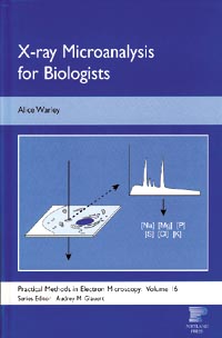
X-ray Microanalysis for Biologists
Alice Warley, 1997, 280pp, softcover,
ISBN 1-855-78055-0.
|